


3D View of Life Processes
Scientists at the Penn State have developed a new liquid-cell technology that enables them to visualize living biological materials and systems in three dimensions under an electron microscope (EM). The technology can help scientists analyze host-pathogen interactions, see a virus is introduced into a cell, and watch molecular mechanisms take place in real-time.
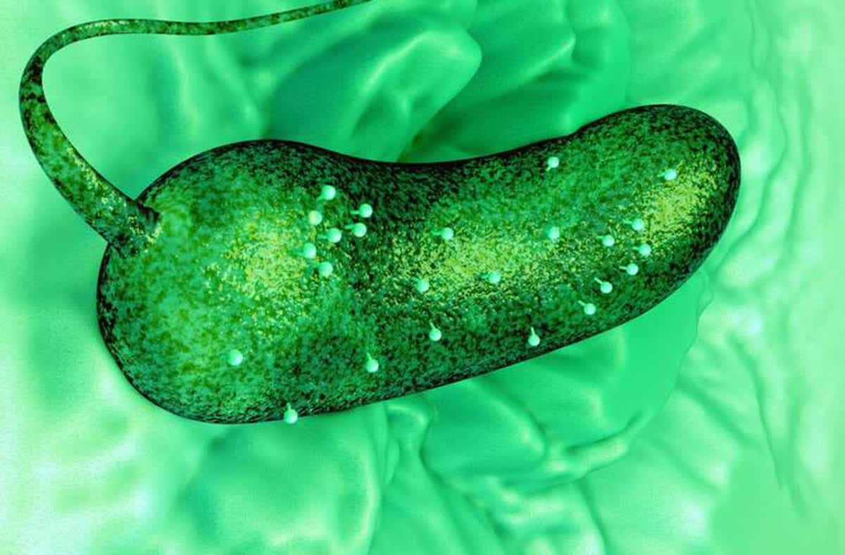
In doing so, scientists demonstrated new insights for the rules of engagement involving a unique bacteriophage and its host bacterium. Their pictures uncovered architectural highlights of the bacteriophage that were previously not well understood.
3D View of Life Processes- The New Technology
Liquid-cell electron microscopy is a rapidly growing field in the imaging domain. While real-time observations are readily available to analyze materials and biological systems, these measurements have been limited to the (2-D) image plane.
William Dearnaley, Lead author and the technical director for Structural Oncology, said that scientists used a copper grid that is coated with a carbon layer and covered that with a silicon nitride chip. A window is situated on the chip, and they pipetted the liquid sample in between the two layers.
This chip design fits into any microscope holder so that it can be universally adapted for
any material.Scientists believe that the new technique- 3D View of Life Processes is expected to be widely adopted in both life sciences and materials science. For example, it can be used in battery research or to look at defects causing building materials to fail.
The study is published in the journal Nano Letters.