Nearly 300,000 new cases of invasive breast cancer are diagnosed every year, of which over 60-75% patients undergo breast-conserving surgery.
The breast conserving surgery known as Lumpectomy aims to remove all cancerous tissues from the breast while leaving behind as much of the healthy tissues as possible, untouched.
But the task is not exactly easy.
In order to identify which cells are cancerous and which are not, a pathology report or a thorough investigation of the cancerous tissue and its surrounding ones is required. But, in comparison to other fields of health, pathology is not all that advanced.
To check if a piece of tissue is cancerous or otherwise, pathologists are still required to go through extensive preparation processes like staining, slicing, and forming tissues into waxy blocks before analyzing them under a microscope. Furthermore, this process can take days, and also limit the number of diagnostic procedures you can carry out on a single sample; since once the tissue is sliced and stained, it can’t be retested for another disease or be used for genetic testing, which is an important part of cancer treatment.
Besides by the time the lab results are back, about 60% of the patients find
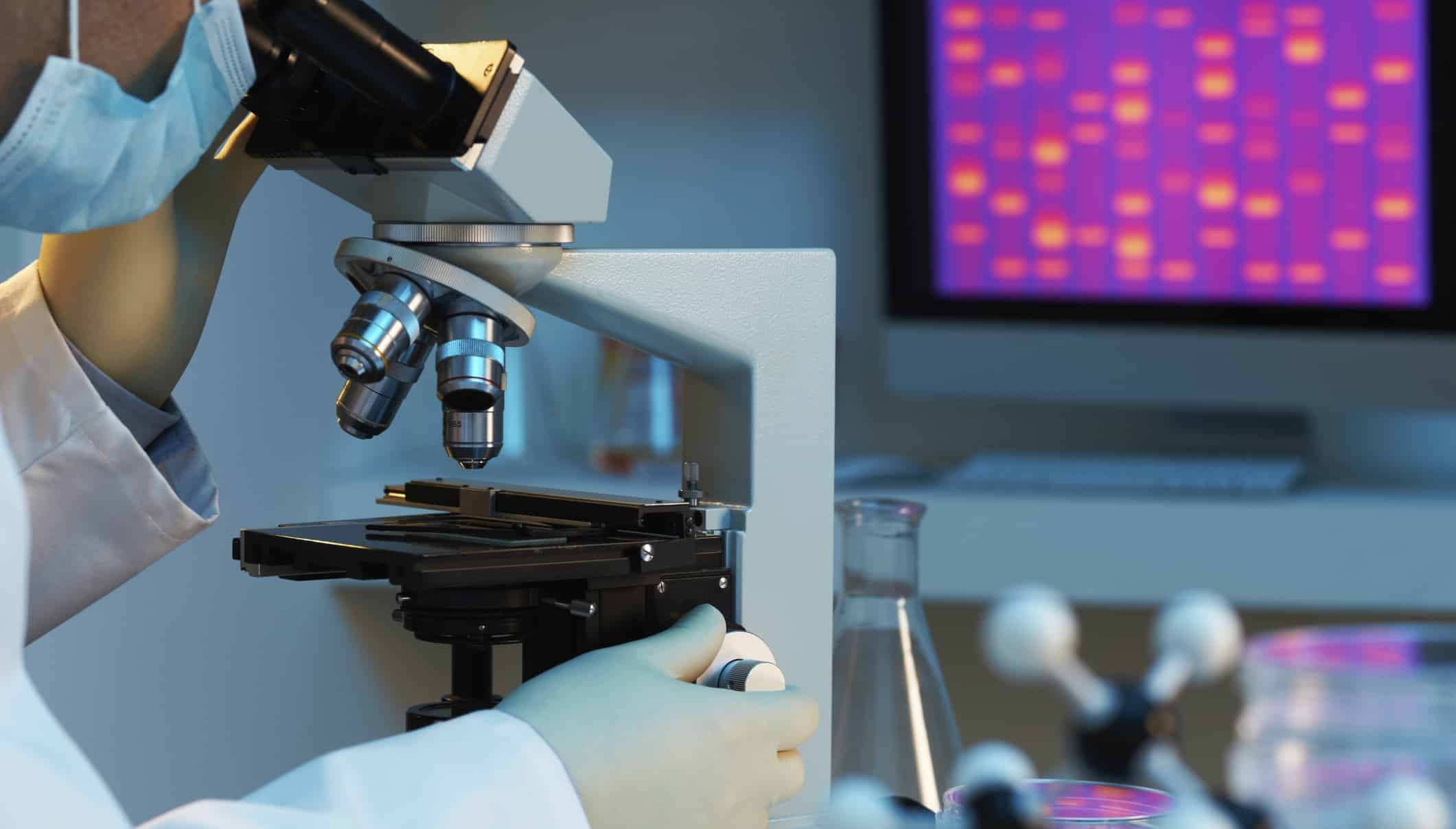
out that they must return for a second surgery to have more tissue removed.“What if we could get rid of the waiting? With 3D photoacoustic microscopy, we could analyze the tumor right in the operating room, and know immediately whether more tissue needs to be removed.” Says Terence T. W. Wang, a Bren Professor of Medical Engineering and Electrical Engineering in Caltech’s Division of Engineering and Applied Science; the lead researcher of the team that has invented a new microscope- 3D photoacoustic microscopy, which is capable of “cutting” through tissue samples using light for the identification of cancerous cells.
The new microscope Photoacoustic microscopy, or simply PAM, excites a tissue sample with a low-energy laser, causing the tissue to vibrate and the system measures the ultrasonic waves emitted by the vibrating tissue. Nuclei usually tend to vibrate more strongly than surrounding material; PAM reveals the size of nuclei and the packing density of cells. Since cancerous tissues have larger nuclei and more densely packed cells, the microscope easily helps us differentiate between the cancerous and normal ones, without requiring tiresome preparation methods.
Researchers say they are currently focusing on breast cancer samples, but in future they plan to use it on cancers that involve solid tumors. Even further into the future, hope to incorporate machine learning algorithms to help pathologists identify cancer more quickly and accurately.


